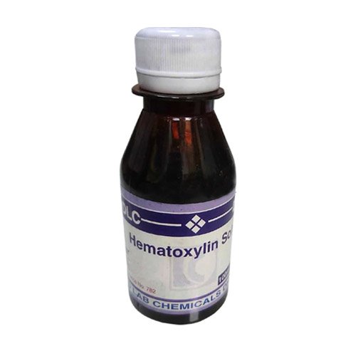
Hematoxylin Solution is a nuclear stain that is used as a counterstain for immunohistochemistry (IHC). Together with eosin, it forms the Hematoxylin and Eosin stain (H&E stain), one of the most commonly used stains in histology.
A common laboratory method uses two dyes called hematoxylin and eosin which make it easier to see different parts of the cell under a microscope. Hematoxylin shows the ribosomes, chromatin (genetic material) within the nucleus, and other structures as a deep blue-purple color.
Hematoxylin is a dye commonly used in histology to stain cell nuclei, making it easier to visualize tissue structures under a microscope. Here’s a basic overview of the hematoxylin solution:
Components of Hematoxylin Solution:
- Hematoxylin: The primary dye derived from the logwood tree.
- Ethanol: Often used as a solvent.
- Water: Typically added to dilute the solution.
- Stabilizers: Sometimes included to enhance shelf life.
Preparation:
- Dissolve Hematoxylin: Mix the hematoxylin powder with ethanol or a suitable solvent.
- Dilute: Add distilled water to achieve the desired concentration (commonly 0.5% to 1%).
- Add Mordant (optional): To enhance staining, a mordant like aluminum sulfate may be included.
Usage of Hematoxylin Solution:
- Staining Procedure:
- Deparaffinize and hydrate the tissue sections.
- Immerse sections in hematoxylin for a specified time (usually a few minutes).
- Rinse with water.
- Differentiate with an acid-alcohol solution if necessary.
- Rinse again and counterstain with eosin if desired.
Storage:
- Store in a dark, cool place to prevent degradation of the dye.
Safety:
- Handle with care, as concentrated solutions can be hazardous. Use gloves and goggles.


Reviews
There are no reviews yet.